


Humoral immunity to adhesins and toxins of the pertussis pathogen in mice immunized with experimental acellular pertussis vaccines from biofilm and planktonic cultures of Bordetella pertussis
- Authors: Zaitsev E.M.1, Britsina M.V.1, Ozerеtskovskaya M.N.1, Zaitsev A.E.1
-
Affiliations:
- I.I. Mechnikov Scientific Research Institute of Vaccines and Sera
- Issue: Vol 102, No 2 (2025)
- Pages: 162-167
- Section: ORIGINAL RESEARCHES
- URL: https://microbiol.crie.ru/jour/article/view/18824
- DOI: https://doi.org/10.36233/0372-9311-631
- EDN: https://elibrary.ru/NQSAYY
- ID: 18824

Cite item

Abstract
Introduction. Whooping cough remains an urgent health problem worldwide, including in countries with high vaccination rates, where, since the 1990s, there has been an increase in the incidence of whooping cough, an increase in the severity of the disease and mortality. In this situation, it is necessary to create a new generation of acellular pertussis vaccines (аPV) that can more effectively affect the colonization, persistence and transmission of Bordetella pertussis. One of the possible directions for improving the vaccine prophylaxis of pertussis infection is the creation of aPV based on protective antigens isolated from biofilm cultures of B. pertussis.
The aim of the study was to research the level of IgG antibodies to the antigens of the pertussis pathogen: adhesins — filamentous hemagglutinin (FHA), pertactin (PRN) and toxins — pertussis toxin (PT), lipopolysaccharide (LPS) in mice immunized with experimental aPV based on antigenic complexes (AC) isolated from biofilm and planktonic cultures of B. pertussis.
Materials and methods. Experimental aPV based on AC isolated from the culture medium of biofilm (aPV-B) and planktonic (aPV-P) cultures of B. pertussis strain No. 317 (serotype 1.2.3) were used in the experiments. IgG titers of antibodies to PT, FHA, PRN and LPS in blood sera of mice immunized with aPV-B and aPV-P was determined in ELISA.
Results. The titers of IgG antibodies to adhesins (FHA and PRN) in the aPV-B group were 8 and 4 times higher, respectively, compared with aPV-P, in the absence of significant differences in the titers of IgG antibodies to PT and LPS.
Conclusion. The higher ability of aPV-B to induce an immune response to B. pertussis adhesins compared to aPV-P, in the absence of significant differences between them in stimulating IgG antibodies to toxins, indicates the advantage of using antigenic complexes from biofilm cultures to create aPV of a new type.
Full Text
Introduction
Pertussis remains a pressing public health problem worldwide, including in countries with a high level of vaccination, where there has been an increase in the incidence of pertussis, an increase in the severity of the disease course and mortality, including among vaccinated children, adolescents and adults since the 1990s [1, 2]. The continued circulation of virulent strains of Bordetella pertussis among the population is associated with the transition from whole-cell vaccines to acellular pertussis vaccines (aPV). aPVs provide protection against severe forms of pertussis, but protective immunity declines rapidly and does not prevent colonization of the respiratory tract and transmission of the pathogen, latent forms of the disease and asymptomatic carriage. Currently known aPVs contain 1 to 5 antigens derived from planktonic cultures of B. pertussis: pertussis toxin (PT) and adhesins: filamentous hemagglutinin (FHA), pertactin (PRN), fimbriae antigens Fim2 and Fim3. One of the probable reasons for the low efficacy of known aPVs is their inability to influence the formation of biofilm forms of B. pertussis in the respiratory tract [3]. The formation of biofilms by B. pertussis strains in the respiratory tract plays an important role in the pathogenesis of pertussis infection by increasing the virulence and persistence of B. pertussis. B. pertussis biofilms differ from planktonic cultures by an altered gene expression spectrum and production of numerous proteins, including adhesins and toxins. In this regard, vaccines from antigens of biofilm and planktonic cultures may differ in immunogenic activity [4].
In this situation, it is necessary to create a new generation of aPVs that can more effectively influence colonization, persistence and transmission of B. pertussis. One of the possible prospects for improving the vaccine prophylaxis of pertussis infection is the creation of aPVs based on protective antigens isolated from biofilm cultures of B. pertussis [5–8].
We have previously shown that the protective activity of aPV-B from biofilm culture was 2.5 times higher than that of aPV-P from planktonic culture during intracerebral infection of mice with a virulent strain of B. pertussis [9]. aPV-B also more effectively reduced the level of colonization by microbial cells of B. pertussis in the lungs of mice during intranasal infection with a virulent strain.
The aim of the study was to investigate the level of IgG antibodies to adhesins and toxins of the pertussis pathogen in mice immunized with experimental aPV based on antigenic complexes isolated from biofilm and planktonic cultures of B. pertussis.
Materials and methods
B. pertussis strain No. 317 (serotype2.3) isolated in Russia from a pertussis patient in 2003, deposited in the Scientific Center for Examination of Medical Devices on 15.09.2017, patent No. 2689903, was used.
F1(CBA×C57Bl6) hybrid mice weighing 12–14 g were obtained from the Andreevka nursery, Moscow region. Animals were kept in vivarium conditions in accordance with the interstate standard for the maintenance and care of laboratory animals (GOST 33217-2014). The authors confirm compliance with institutional and national standards for the use of laboratory animals in accordance with the “Consensus Author Guidelines for Animal Use” (IAVES, 23.07.2010). The study protocol was approved by the Ethical Committee of the I.I. Mechnikov Research Institute (protocol No. 15, December 25, 2024)
Control of morphologic, serologic and culture properties of B. pertussis strain No. 317 was carried out in accordance with the Methodological Instructions. Cultivation of the strain in liquid synthetic nutrient medium, isolation of antigenic complexes (AC) from planktonic and biofilm cultures was carried out in accordance with the previously described method [10]. To characterize the composition of AC from biofilm and planktonic cultures, vertical electrophoresis in polyacrylamide gel (PAAG) under denaturing conditions according to Lammli [11] was used. Electrophoresis was performed in 10% Tris-glycine buffer at a current of 25 mA. At the end of the process, the gel was stained with Coomassie brilliant blue R-250, after which it was washed twice in an aqueous solution containing 10% acetic acid and 35% ethanol.
Detoxification of B. рertussis antigenic complexes was carried out with formalin to a concentration of 0.4% with the addition of sucrose (10%) for 20 days with periodic shaking at 37.0 ± 0.5°C. To obtain aPV, antigenic complexes were sorbed on 2% aluminum hydroxide gel (InvivoGen) in such a ratio that 1 ml of the mixture contained 50 μg of protein, 0.3 mg of aluminum hydroxide and PBS to 1 ml [10]. To study the level and dynamics of IgG antibodies to PT, PRN, FHA and lipopolysaccharide (LPS) (all from National Institute for Biological Standards and Control), mice of line F1(CBA×C57BL6) weighing 12–14 g were immunized intraperitoneally (n = 20 in each group) three times with an interval of 7 days with experimental aPV at a dose of 25 μg. Blood was drawn from mice at 7, 14, 21 and 28 days after the last immunization. Blood sampling (total) from mice was performed under ether anesthesia.
The level of IgG antibodies in the sera of immunized mice was detected by enzyme-linked immunosorbent assay. Sera of intact mice (n = 5) were used as a negative control. The concentration of antigens for adsorption on the plates was: PT — 2 μg/mL; FHA — 2 μg/mL; PRN — 2 μg/mL; LPS — 2.5 μg/mL. Peroxidase antispecies conjugate to mouse IgG (Invitrogen) and tetramethylbenzidine as substrate mixture were used in the experiments. The reaction results were counted using a Multiskan vertical scanning spectrophotometer (Thermo Scientific) at a wavelength of 450 nm. The values inverse to their maximum dilutions, at which the optical density (OD) values were 2 or more times higher than the OD values in the negative control wells, were taken as the serum titer.
Statistical analysis was performed using the Microsoft Office Excel application program package. Quantitative data are presented as M ± m. Comparisons were performed using Student's t test. Differences were considered significant at p < 0.05.
Results
From the culture medium of biofilm and planktonic cultures of strain No. 317, ACs were isolated, which were used as the basis for the production of two variants of aPV: aPV-B and aPV-P.

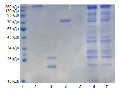
Fig. 1. Electrophoresis in PAAG from antigenic complexes of biofilm and planktonic cultures of B. pertussis.
The tracks on the electrophoregram are: 1 — molecular weight markers; 2 — FHA; 3 — PT; 4 — PRN; 5 — LPS; 6 — AC strain No. 317 (biofilm culture); 7 — AC strain No. 317 (planktonic culture).
The results of analysis of crude and not adsorbed on aluminum hydroxide gel ACs isolated from biofilm and planktonic cultures of strain No. 317 by electrophoresis in PAAG are presented in Fig. 1. Proteins in the range of molecular masses from 15 to 220–250 kDa were detected in the AC composition of strain No. 317. At the same time, the intensity of protein bands with a molecular mass of 15 kDa was higher in AC from biofilm culture compared to AC from planktonic culture. FHA (220 kDa), PRN (69 kDa), and proteins with molecular masses of about 28 kDa, less than 26 kDa, and greater than 15 kDa corresponding to PT fragments were detected in both preparations. No protein components were detected on the LPS lane, indicating the immunochemical purity of the preparation used.

Fig. 2. Maximum titers of IgG antibodies to B. pertussis antigens in mice immunized with aPV-B and aPV-P.
On the Y-axis: antibody titers. On the X-axis: 1 — PT aPV-P; 2 — PT aPV-B; 3 — FHA aPV-P; 4 — FHA aPV-B; 5 — PRN aPV-P; 6 — PRN aPV-B; 7 — LPS aPV-P; 8 — LPS aPV-B. IgG antibody titers in intact mice < 100.
The results of IgG antibody levels to PT, PRN, FHA and LPS in mice immunized with aPV from biofilm and planktonic cultures of B. pertussis are shown in Fig. 2. The titers of IgG antibodies to PT in both groups reached maximum values on the 21st day with a subsequent decrease on the 28th day. Differences in IgG antibody titers to PT in groups aPV-B and aPV-P were statistically insignificant. The maximum titers of IgG antibodies to PRN in the aPV-B group were observed on the 14th and 21st days, and in the aPV-P group — on the 14th day with subsequent decrease in both groups. The titers of IgG antibodies to PRN in the aPV-B group were significantly higher than in the aPV-P group. The titers of IgG antibodies to FHA in aPV-B and aPV-P groups consistently increased and reached the maximum values on the 21st day. Further, a decrease in IgG antibody titers was noted on the 28th day. The maximum titers of IgG antibodies to FHA and PRN in the aPV-B group were 8 and 4 times higher than in the aPV-P group, respectively. The maximum titers of IgG antibodies to LPS in the aPV-B group were observed on the 14th day, and in the aPV-P group – on the 21st day with subsequent decrease. Differences in IgG antibody titers to LPS in both groups were statistically unreliable.
Discussion
B. pertussis produces a number of virulent factors that determine the pathogenetic mechanism of pertussis infection. They can be divided into adhesins (fimbriae, PRN, tracheal colonization factor, FHA) and toxins (PT, adenylate cyclase, tracheal cytotoxin, dermonecrotic toxin, LPS (endotoxin)). The adhesins provide fixation of the pathogen on the epithelial cells of the respiratory tract, and the toxins have a direct damaging effect. The main adhesin of B. pertussis is FHA, which is a protein with a molecular mass of 220 kDa, not associated with fimbriae [12]. PRN is a non-fimbrial protein (69 kDa) associated with the outer membrane of the microbial cell. PRN has no toxic properties and is an adhesin by its pathogenetic action and has immunomodulatory activity [13, 14].
PT is one of the main pathogenicity factors of B. pertussis, causes various biological effects in vivo and in vitro and accounts for a significant part of the disease symptoms in pertussis patients. PT is an exotoxin secreted by the microbial cell and is a protein with a molecular mass of 117 kDa, consisting of 5 structural units (S1, S2, S3, S4 and S5) with molecular masses ranging from 28 kDa for S1 to 9.3 kDa for S5 [15, 16].
LPS is a component of the outer part of the cell membrane of all Gram-negative bacteria, including B. pertussis. LPS molecules ensure the structural integrity of the bacterial cell and protect the membrane from aggressive environmental influences. Side effects of whole-cell pertussis vaccines are predominantly associated with B. pertussis LPS [17, 18].
Due to genotypic and phenotypic polymorphism, as well as depending on the culture conditions (biofilm or planktonic cultures), B. pertussis strains may differ in the levels of production of PT, FHA, PRN and other antigens. B. pertussis biofilms are formed as a result of complex coordinated interactions between microbial cells and biotic and abiotic substrates. In biofilm cultures, the expression of adhesins increases, which promotes attachment to the substrate and intercellular interactions.
We investigated the composition of B. pertussis planktonic and biofilm cultures and the level of IgG antibodies to adhesins (PRN, FHA) and PT and LPS toxins (endotoxin) in mice immunized with aPV from planktonic and biofilm cultures of B. pertussis strain No. 317. According to the data of electrophoresis in PAGE, the studied ACs had in their composition FHA, PRN and PT fragments — the main protective antigens of B. pertussis, which are part of aPV. In general, the electrophoregrams of both preparations were almost identical, except for the greater intensity of protein bands with a molecular mass of about 15 kDa in aPV-B. Increasing titers of IgG antibodies to PT, FHA and PRN were found in the sera of mice immunized with both preparations, which confirms the results of electrophoresis about the presence of these antigens in the composition of aPV-B. The dynamics of IgG antibody titers to PT, FHA, PRN and LPS in aPV-B and aPV-P groups in general had a similar character with the antibody titers increasing, reaching maximum values and subsequent decrease. At the same time, significant differences between aPV-B and aPV-P in the levels of antibodies to FHA and PRN adhesins were detected. The antibody titers to FHA and PRN in the aPV-B group were significantly higher than in the aPV-P group, which can be explained by the different specific content of these antigens in the composition of aPV due to a higher level of adhesin production by the biofilm culture or higher immunogenicity of FHA and PRN in the composition of aPV-B. There were no significant differences between aPV-B and aPV-P in antibody titers to PT and LPS.
Conclusion
The higher ability of aPV-B to induce an immune response to B. pertussis adhesins, which provide fixation of the pathogen on respiratory tract epithelial cells, compared to aPV-P, with no significant differences between both preparations in stimulation of IgG antibodies to PT, indicates the advantage of using antigenic complexes from biofilm cultures to create more immunogenic aPV of a new type.
About the authors
Evgeny M. Zaitsev
I.I. Mechnikov Scientific Research Institute of Vaccines and Sera
Author for correspondence.
Email: lab.immunomod@yandex.ru
ORCID iD: 0000-0002-4813-9074
D. (Med.), Head, Laboratory of immunomodulators
Russian Federation, MoscowMarina V. Britsina
I.I. Mechnikov Scientific Research Institute of Vaccines and Sera
Email: britsinamarina@yandex.ru
ORCID iD: 0000-0002-3044-0790
Cand. Sci. (Biol.), leading researcher, Laboratory of immunomodulators
Russian Federation, MoscowMaria N. Ozerеtskovskaya
I.I. Mechnikov Scientific Research Institute of Vaccines and Sera
Email: manja33@yandex.ru
ORCID iD: 0000-0001-9809-4217
Cand. Sci. (Med.), leading researcher, Laboratory of immunomodulators
Russian Federation, MoscowAnton E. Zaitsev
I.I. Mechnikov Scientific Research Institute of Vaccines and Sera
Email: anton.zajtseff2015@yandex.ru
ORCID iD: 0000-0002-8434-231X
Cand. Sci. (Med.), researcher, Laboratory of therapeutic vaccines
Russian Federation, MoscowReferences
- Ломоносова А.В. Причины и последствия несвоевременной вакцинации против коклюшной инфекции в Российской Федерации. Журнал микробиологии, эпидемиологии и иммунобиологии. 2020;97(5):492–502. Lomonosova A.V. Causes and consequences of delayed vaccination against pertussis infection in the Russian Federation. Journal of Microbiology, Epidemiology and Immunobiology. 2020;97(5):492–502. DOI: https://doi.org/10.36233/0372-9311-2020-97-5-11, EDN: https://elibrary.ru/pdbbte
- Stefanelli P. Pertussis: identification, prevention and control. Adv. Exp. Med. Biol. 2019;1183:127–36. DOI: https://doi.org/10.1007/5584_2019_408
- Fullen A.R., Gutierrez-Ferman J.L., Yount K.S., et al. Bps polysaccharide of Bordetella pertussis resists antimicrobial peptides by functioning as a dual surface shield and decoy and converts Escherichia coli into a respiratory pathogen. PLoS Pathog. 2022;18(8):e1010764. DOI: https://doi.org/10.1371/journal.ppat.1010764
- Suyama H., Luu L.D.W., Zhong L., et al. Integrating proteomic data with metabolic modeling provides insight into key pathways of Bordetella pertussis biofilms. Front. Microbiol. 2023;14:1169870. DOI: https://doi.org/10.3389/fmicb.2023.1169870
- Fullen A.R., Gutierrez-Ferman J.L., Yount R.S., et al. Bps polysaccharide of Bordetella pertussis resists antimicrobial peptides by functioning as a dual surface shield and decoy and converts Escherichia coli into a respiratory pathogen. PLoS Pathog. 2022;18(8):e1010764. DOI: https://doi.org/10.1371/journal.ppat.1010764
- Fullen A.R., Gutierrez-Ferman J.L., Rayner R.E., et al. Architecture and matrix assembly determinants of Bordetella pertussis biofilms on primary human airway epithelium. PLoS Pathog. 2023;19(2):e1011193. DOI: https://doi.org/10.1371/journal.ppat.1011193
- Carriquiriborde F., Martin Aispuro P., Ambrosis N., et al. Pertussis vaccine candidate based on outer membrane vesicles derived from biofilm culture. Front. Immunol. 2021;12:730434. DOI: https://doi.org/10.3389/fimmu.2021.730434
- Dorji D., Graham R.M., Singh A.K., et al. Immunogenicity and protective potential of Bordetella pertussis biofilm and its associated antigens in a murine model. Cell. Immunol. 2019;337: 42–7. DOI: https://doi.org/10.1016/j.cellimm.2019.01.006
- Zaytsev E.M., Britsina M.V., Ozeretskovskaya M.N., Zaitsev A.E. Protective activity and safety of experimental acellular pertussis vaccines based on antigenic complexes isolated from biofilm and planktonic cultures of Bordetella pertussis. Bull. Exp. Biol. Med. 2024;177(3):349–52. DOI: https://doi.org/10.1007/s10517-024-06187-9
- Зайцев Е.М., Бажанова И.Г., Брицина М.В. и др. Бесклеточная коклюшная вакцина из антигенов свежевыделенного штамма B. pertussis серовара 1.2.3. Журнал микробиологии, эпидемиологии и иммунобиологии. 2020;97(2):134–9. Zaitsev E.M., Bazhanova I.G., Britsina M.V., et al. Cell-free pertussis vaccine from antigens of freshly isolated strain of B. pertussis serotype 1.2.3. Journal of Microbiology, Epidemiology and Immunobiology. 2020;97(2):134–9. DOI: https://doi.org/10.36233/0372-9311-2020-97-2-134-139 EDN: https://elibrary.ru/cqzssv
- Laemmli U.K. Cleavage of structural proteins during the assembly of the head of bacteriophage T4. Nature. 1970; 227(5259):680–5. DOI: https://doi.org/10.1038/227680a0
- Imani D., Bahadori T., Golsaz-Shirazi F., et al. High purity and recovery of native filamentous hemagglutinin (FHA) from Bordetella pertussis using affinity chromatography. J. Chromatogr. B Analyt. Technol. Biomed. Life Sci. 2024;1239:124122. DOI: https://doi.org/10.1016/j.jchromb.2024.124122
- Imani D., Bahadori T., Ghourchian S., et al. Novel mouse monoclonal antibodies against Bordetella pertussis pertactin antigen with versatile applications. J. Microbiol. Methods. 2023;211:106786. DOI: https://doi.org/10.1016/j.mimet.2023.106786
- Silva R.P., DiVenere A.M., Amengor D., Maynard J.A. Antibodies binding diverse pertactin epitopes protect mice from Bordetella pertussis infection. J. Biol. Chem. 2022;298(3):101715. DOI: https://doi.org/10.1016/j.jbc.2022.101715
- Scanlon K., Skerry C., Carbonetti N. Association of pertussis toxin with severe pertussis disease. Toxins (Basel). 2019;11(7):373. DOI: https://doi.org/10.3390/toxins11070373
- Locht C., Antoine R. The history of pertussis toxin. Toxins (Basel). 2021;13(9):623. DOI: https://doi.org/10.3390/toxins13090623
- Locht C. Pasteurian contributions to the study of Bordetella pertussis toxins. Toxins (Basel). 2023;15(3):176. DOI: https://doi.org/10.3390/toxins15030176
- Koj S., Ługowski C., Niedziela T. Bordetella pertussis lipooligosaccharide-derived neoglycoconjugates — new components of pertussis vaccine. Postepy. Hig. Med. Dosw. (Online). 2015;69:1013–30. (in Polish)
Supplementary files

