


Development and evaluation of a recombinant monoclonal human antibody with virus-neutralizing activity against the F glycoprotein of respiratory syncytial virus
- Authors: Klotchenko S.A.1, Romanovskaya-Romanko E.A.1, Plotnikova M.A.1, Pulkina A.A.1, Shaldzhyan A.A.1, Balabashin D.S.2, Toporova V.A.1,2, Aliev T.K.2,3, Gyulikhandanova N.E.1, Lioznov D.A.1,4
-
Affiliations:
- Smorodintsev Research Institute of Influenza
- Shemyakin–Ovchinnikov Institute of bioorganic chemistry
- Lomonosov Moscow State University
- First St. Petersburg State Medical University named after Academician I.P. Pavlov
- Issue: Vol 101, No 6 (2024)
- Pages: 735-747
- Section: ORIGINAL RESEARCHES
- URL: https://microbiol.crie.ru/jour/article/view/18697
- DOI: https://doi.org/10.36233/0372-9311-611
- EDN: https://elibrary.ru/zkqvtw
- ID: 18697

Cite item

Abstract
Introduction. Respiratory syncytial virus (RSV) is the most common pathogen causing lower respiratory tract infections in children. RSV also poses a serious threat to the elderly and immunocompromised patients. Developing a therapy based on recombinant human antibodies to block the RSV fusion (F) glycoprotein is urgent to reduce the incidence of RSV infections and prevent associated complications.
Aim. To design plasmid vectors for efficient production of the recombinant monoclonal antibody FM1 in a eukaryotic expression system targeting the RSV fusion (F) glycoprotein and to evaluate its activity against RSV subtypes A and B in vitro.
Materials and methods. Constructs encoding the recombinant antibody FM1 were designed using genetic engineering. Recombinant antibodies were produced in the CHO-K1 cell line through transient expression. Antibody specimens were purified from the culture supernatant using affinity chromatography, with a modified protein A as the ligand. The virus-neutralizing activity of the antibody was evaluated in a microneutralization assay using several RSV strains on a Vero cell monolayer culture.
Results. We developed a two-plasmid vector system to produce the recombinant FM1 antibody targeting the RSV F glycoprotein, using CHO cells as transient producers. The antibody was successfully produced, purified, and characterized, with its biological activity confirmed. The FM1 antibody demonstrated enhanced virus-neutralizing activity against reference and seasonal RSV strains of subtypes A and B compared to the control drug palivizumab.
Conclusion. A recombinant FM1 antibody-based drug could address the import substitution challenge for protective measures against RSV infection. The authors are currently developing a stable FM1 producer clone with high productivity and viability and investigating the therapeutic efficacy of this antibody in a sublethal RSV infection mouse model.
Full Text
Introduction
Respiratory syncytial virus (RSV) is the most prominent pathogen of lower respiratory tract infections in children and also poses a serious threat to the elderly and immunocompromised patients [1, 2].
Up to 70% of children have their first RSV infection (RSVI) before the age of 1 year, and almost every child is infected during the first 3 years of life. The frequency of RSVI verification in children under 3 years of age hospitalized for lower respiratory tract infection reaches 42–63% in developed countries [3, 4]. Bronchiolitis is the most common (50–90%), pneumonia (5–40%) and tracheobronchitis (10–30%) are slightly less common, and mortality averages 1% [5–7]. According to the results of meta-analysis of morbidity in 132 developed countries, RSVI accounts for more than 3 million hospitalizations per year and about 60 thousand deaths among children under 5 years of age [8, 9].
According to the data of polymerase chain reaction diagnostics conducted at the A.A. Smorodintsev Research Institute of Influenza, in the season of 2023–2024, during the peak period of acute respiratory viral infections the share of RSV among respiratory pathogens amounted to 26% excluding SARS-CoV-2 and influenza viruses and 16% including influenza viruses, which clearly indicates a significant role of RSVI in the structure of respiratory infections, especially in children under 2 years of age. According to the data of the A.A. Smorodintsev Research Institute of Influenza, the share of RSVI among hospitalized patients is 13-19% [10]. Given that about 30 million patients with respiratory infections are registered annually in Russia, RSVI accounts for at least 3.9 million of these cases.
According to the estimates of T. Shi et al., 45% of hospitalizations and in-hospital deaths in children under 6 months of age are due to acute respiratory failure resulting from RSVI [9]. Since vaccines are less immunogenic at this age, maternal immunization or monoclonal antibody (MCA) administration can be used to induce passive immunity in infants to provide better protection for the child.
Antibodies are thought to play a key role in limiting acute lower respiratory tract infection in RSVI. Recent studies emphasize that induction of mucosal immunity is required for a full immune response to protect against reinfection [11]. Until recently, the only means of preventing RSVI was the humanized MCA palivizumab [12], which was used only in at-risk groups and required repeated injections. Currently, a new drug, nirsevimab, has been developed and approved in the EU and the USA, which is more stable and can be administered once [13–15]. In Russia, only palivizumab is registered for clinical use. The development of a preparation for the prevention and therapy of RSVI based on recombinant human neutralizing antibodies interacting with the surface glycoprotein F of RSVI will significantly reduce the incidence of RSVI in young children, reduce disability and mortality caused by this pathogen, and prevent the development of complications of this infection. There is also an urgent need for means of prevention and therapy of RSVI in the elderly and immunocompromised patients.
The aim of the present study was to construct plasmid vectors for the accumulation of highly active recombinant MCA (rMCA) FM1 in a eukaryotic expression system directed against the glycoprotein F of RSV and to evaluate the specific activity of the resulting antibody against different strains of RSV subtypes A and B in vitro.
Materials and methods
Construction of plasmid vectors
Nucleotide insertions encoding the heavy and light chains of rMCA FM1 (including constant sites) were assembled on the basis of published sequences of the MEDI8897 antibody [16] and synthesized at Eurogen. Cloning was performed into pVAX1 vector using restriction sites for Nhe I and Xho I endonucleases. Colonies were screened by polymerase chain reaction, the presence of target insertions was confirmed by Sanger sequencing at Eurogen, after which the created plasmid constructs pVAX1-FM1-HC and pVAX1-FM1-LC were accumulated, purified using the Plasmid Miniprep 2.0 kit (Eurogen) and used for transfection of eukaryotic cells.
DNA electrophoresis in agarose gel
Plasmid DNA preparations and amplicons were analyzed in 0.8% agarose gel prepared in 1× TAE buffer containing up to 0.5 μg/ml ethidium bromide using 6× DNA plating buffer. The results of electrophoretic separation were visualized using a Gel Doc EZ Imager (Bio-Rad).
Cells and viruses
CHO-K1 cells (Chinese hamster ovary cells, ATCC #CCL-61) and Vero cells (African green monkey kidney cells, ATCC #CCL-81) obtained from the ATCC cell culture bank (American Type Culture Collection) were used in the experiments. CHO-K1 cells were cultured on F12K medium (Gibco) supplemented with 10% FBS (Gibco), while Vero cells were cultured on α-MEM medium (Biolot) supplemented with 5% FBS (Gibco). All experiments (except for the stages of selection of CHO-K1 producers) were performed without antibiotics. Daily cultures were used in this study. All cell cultures were maintained at a temperature of 37.0 ± 0.5ºC, in an atmosphere of 5% CO2, under high humidity conditions (80-100%).
We used RSV of two reference strains: A2 (subtype A), infectious titer 7.7 lgTID50/mL and 9320 (subtype B), infectious titer 6.8 lgTID50/mL; as well as 2 seasonal RSV isolates: hRSV/A/Russia/RII-26062v/2022 (subtype A), infectious titer 6.8 lgTID50/mL and hRSV/B/Russia/RII-4759/2022 (subtype B), infectious titer 6.3 lgTID50/mL (Collection of the A.A. Smorodintsev Research Institute of Influenza).
The commercial drug Synagis (AstraZeneca; solution for intramuscular administration, 100 mg/ml, series 406039, manufactured 08.2023, valid until 07.2026), which is a humanized MCA palivizumab directed against glycoprotein F of RSV, was used as a comparison preparation [17].
Enzyme immunoassay
Enzyme-linked immunosorbent assay (ELISA) in sandwich format was performed using 96-well Microlon High Binding plates (Greiner Bio-One), PST-60HL-4 thermoshaker (BioSan), commercial MCAs, control preparation of palivizumab, and recombinant antibodies and purified viruses obtained at the A.A. Smorodintsev Research Institute of Influenza. Capture antibodies against Fc-fragments of human immunoglobulin heavy chains (#ab77118, Abcam) were sorbed at a concentration of 1 μg/mL in a volume of 100 μl per well at 4ºC overnight. Blocking was performed with a solution of 5% skim milk (Blotting-Grade Blocker, #1706404, Bio-Rad) on PBST (Tween-20 to 0.05%) at 37ºC for 1 h. Incubation with the analyzed samples was performed at 37ºC for 2 h, after which detection antibodies against human immunoglobulin light kappa chains (#4G7cc, Hytest) conjugated to horseradish peroxidase were added at the manufacturer's recommended concentration and incubated at 37ºC for 1 h. After standard detection using a substrate mixture of tetramethylbenzidine (Chema) and mononormal sulfuric acid, optical density (OD) was measured at wavelengths of 450 nm (OD450) and 655 nm (OD655) on a Multiskan SkyHigh microplate spectrophotometer (Thermo Fisher Scientific). The average value of OD450–655 for all negative controls plus 3 standard deviations was taken as the threshold value.
Chromatographic purification of recombinant antibodies
Chromatographic purification of recombinant antibodies was performed by affinity chromatography using an AKTA pure chromatography system on a 5 mL MabPurix P45 column (Sepax). The column was washed with 10 CV (column volume) of starting buffer (1× PBS) at a flow rate of 5 mL/min. The culture liquid (50 mL), pre-filtered through a Sartorius syringe filter (pore size 0.45 μm, polyethersulfone membrane material), was introduced into the chromatograph through a sample pump at a flow rate of 2.5 mL/min. The column was then washed with 10 CV of starting buffer at a flow rate of 5 mL/min. Antibodies were eluted with 100% elution buffer (20 mM glycine, pH 3.0) in a volume of 15 CV at a flow rate of 5 mL/min. Monitoring was performed using OD280. During the elution step, peaks with OD above 0.05 AU were selected using an automated collector. To the collected material, 1 M Tris-HCl pH 8.8 solution (20 μL/mL) and 4 M NaCl solution (40 μL/mL) were added. To increase sterility and prevent degradation and contamination, the obtained preparation was filtered using a Sartorius syringe filter (pore size 0.45 μm, polyethersulfone membrane material) and used for further studies.
Protein electrophoresis in polyacrylamide gel
Polyacrylamide gel (PAAG) electrophoresis was performed according to the Lammlie method [18] under reducing (in the presence of β-mercaptoethanol) and non-reducing conditions. A 15-well Any kD gradient gel (#4568126, Bio-Rad) was used. The sample was mixed with 4-fold Lammlie's buffer before being applied to the PAAG wells, followed by protein denaturation at 95ºC in a Gnome solid-state thermostat (DNA-Technology) for 10 min (reducing conditions). 2.5 μg of protein sample was added to each well. Protein concentration was evaluated on a NanoDrop ND-1000 spectrophotometer (Thermo Fisher Scientific), and for antibodies the IgG mode was used, with an E value of 1% = 13.70. Electrophoretic separation of proteins was performed at constant current (25 mA per gel) for 45 min in a Mini-PROTEAN Tetra vertical electrophoretic cell (Bio-Rad). The gel was stained with Coomassie colloidal solution [19]. The stained gel was imaged on a Gel Doc EZ Imager gel documentation station (Bio-Rad).
Microneutralization reaction and determination of half-maximal inhibitory dose
The virus-neutralizing activity of recombinant antibodies was evaluated on Vero monolayer cell culture using the method described previously [20]. A series of triplicate dilutions of recombinant antibody preparations (3 independent repeats) were mixed with an equivalent volume of growth medium containing 100 TID50 RSV, and after 1 h incubation at room temperature, the dilutions were transferred to plates with a daily monolayer of Vero cells. The plates were incubated for 4 days in a CO2 incubator at 37.0 ± 0.5ºC under high humidity conditions (80–100%). Inhibition of RSV replication in the presence of different concentrations of rMCA was determined on the 4th day after infection by cell ELISA using as primary mouse antibody MCA 4F2 specific to RSV F-glycoprotein of subtypes A and B (A.A. Smorodintsev Research Institute of Influenza) and horseradish peroxidase-labeled goat anti-mouse IgG (H+L) secondary antibody (Bio-Rad). After conjugate-substrate color reaction, the OD was measured using a Multiskan SkyHigh microplate spectrophotometer (Thermo Fisher Scientific) and calculated as the difference of OD450–620. The obtained OD values were transformed into the percentage of inhibition of the development of cytopathic action of the virus at a certain concentration of recombinant antibodies. The half-maximal inhibitory concentration (IC50) was calculated by plotting a four-parameter dose-effect curve using GraphPad Prism 9.5.1 software on the basis of 3 independent repeats.
Primary data and statistical processing
Statistical analysis of primary data was performed in the Microsoft Office Excel 2010 and GraphPad Prism 9.5.1 software packages. The following statistical indicators were used to present the data: standard deviation, arithmetic mean, standard error of the mean. The Shapiro–Wilk test was used to test the hypothesis of normality of the obtained distribution of values, and Student's t-test was used to determine the significance of differences between group averages. The a priori level of significance was taken as α = 0.05. Differences were considered reliable at the achieved significance level p < α [21].
Results
Design and generation of expression constructs encoding recombinant FM1 antibody
The sequences of the MEDI8897 antibody were chosen as the basis for the design of a long-acting human rMCA FM1 for the prevention of lower respiratory tract diseases caused by RSVI [16].
The antibody under development is a recombinant human immunoglobulin of the IgG1κ class. MEDI8897 has the following sequences of hypervariable regions: in the light chain, L1 QASQDIVNYLN, L2 VASNLET and L3 QQYDNLPLT; in the heavy chain, H1 DYIIN, H2 GIIPVLGTVHYGPKFQG and H3 VSETYLPHYFDN [16]. The constant region of the MEDI8897 light chain belongs to the human κ-isotype (encoded by the κ locus 2p11.2 on the 2nd chromosome) and is completely identical to the canonical sequence P01834 presented in the UniProt open protein sequence database [22]. The constant sequence of the heavy chain MEDI8897 belongs to the immunoglobulin G1 class (sIgG1, secreted form) and has several differences from the sequence P01857 presented in UniProt. In particular, in addition to the intentionally introduced 3 amino acid substitutions in the CH2 domain of the constant region (M257Y/S259T/T261E, [YTE]) in MEDI8897 that ensure prolonged circulation of the antibody in the blood [16], we also detected 2 substitutions in the constant regions of MEDI8897: K97R (VAR_003886) and D239E (VAR_003887), which are variant natural substitutions in the alleles [22]. Thus, despite the presence of variation, the antibody amino acid sequences that fully match the heavy and light chain sequences of MEDI8897 were selected for cloning [16].
A 2-plasmid expression system was chosen for FM1 rMCA production, which implies the presence of 2 vector constructs, one of which encodes the heavy and the other the light full-length chains of the antibody, i. e. the chains in the constructs contain both variable and constant regions of the antibody, as well as signal peptides.
The pVAX1 vector containing CMV promoter, T7-promoter at the 5′-end of the insert and polyadenylation site (from bovine growth hormone) was selected for cloning; selective antibiotics for the vector were: in bacterial system — ampicillin, in eukaryotic system — neomycin (kanamycin).
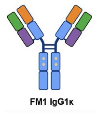
The pVAX1-based pVAX1-FM1-HC (total length 4349 bp) and pVAX1-FM1-LC (3632 bp) constructs capable of expression and production of FM1 rMCA in eukaryotic cells in the format of a full-length IgG1κ heterotetramer were assembled by genetic engineering methods. Schematic representations of the resulting constructs are shown in Fig. 1. Both sequences, the heavy chain (total length 1428 bp, variable part size 378 bp) and the light chain (total length 711 bp, variable part size 321 bp), contain the Kozak sequence, N-terminal leader peptides that ensure secretion of the full-length antibody, and are flanked by restriction sites Nhe I (at the 5′-end) and Xho I (at the 3′-end).

Fig. 1. Design and production of expression constructs encoding the recombinant antibody FM1 in the format of a full-size IgG1κ heterotetramer.
FM1-HC — heavy chain, FM1-LC — light chain, SP — signaling peptides, VH-FM1 — variable domain of heavy chain, CH1, CH2 and CH3 — constant domains of heavy chain, hinge — hinge section, VL-FM1 — variable domain of light chain, CL-kappa — constant domain of the light κ-chain.
The developed plasmid constructs are capable of constitutive expression in eukaryotic cells (due to the presence of the CMV promoter) of mature polyadenylated mRNAs encoding the heavy and light chains of rMCA FM1.
Production of stable eukaryotic pools of recombinant FM1 antibody by temporary transfection
FM1 rMCA production was carried out by temporary transfection of eukaryotic cell line CHO-K1 with a 2-plasmid system providing the correct conformation and correct glycosylation of the formed antibody. To accumulate FM1 rMCA, we chose the monoselection variant, in which CHO-K1 cells were transfected with plasmid constructs based on a single pVAX1 vector. For this purpose, the obtained constructs pVAX1-FM1-HC and pVAX1-FM1-LC and commercial reagent Lipofectamine 3000 (Thermo Fisher Scientific) were used. The antibiotic geneticin (an analog of neomycin) in the concentration range of 100-400 μg/mL (for selection of clones carrying the NeoR/KanR resistance gene) was used as a selective agent for progenitor cells obtained using the CHO-K1 line.
To increase the probability of obtaining a greater number of antibody-producing clones, adaptation of the pool of transfected cells to selective conditions was performed before each cloning. The adaptation process consisted of passaging cells on selective medium every 3–4 days. At this time, the cells deprived of the genetic construct, including the selective marker gene, died. After several passages, the viability of cells in the pool was restored due to an increase in the growth rate of cells adapted to the selective medium.
According to the results of sandwich ELISA (using lower antibodies against Fc-fragment of heavy chains and upper antibodies against light κ-chains of human immunoglobulins, allowing to detect only full-size IgG1κ heterotetramers) transfected CHO-K1 cells were capable of stable production of FM1 antibody. The dynamics of rMCA accumulation was assessed during the first 6 days, thereafter the cells formed a 100% monolayer, the production of the target product by the cells reached a constant level and directly correlated with the number of cells. The concentration of FM1 rMCA in supernatants obtained from CHO-K1 cells during transient expression was measured by sandwich ELISA and was 10 μg/mL. After 15 days of culturing, it was possible to obtain populations capable of stable propagation under selection conditions and producing FM1 rMCA. Further, the obtained temporary CHO-producers were used for accumulation of preparative amount of rMCA FM1 and its subsequent chromatographic purification.
Accumulation, purification, integrity analysis and evaluation of specific activity of recombinant FM1 antibody
FM1 rMCA was purified from the culture fluid by affinity chromatography using modified protein A (MabPurix P45, Sepax) as a ligand. Supernatant from CHO-K1 cells was collected for 1 month every 5 days. Purification was performed from approximately 300 ml of cell supernatant to yield 2.8 mg of FM1 rMCA preparation for studies of its specific and virus-neutralizing activity.
The chromatogram of the purification of the original preparation by affinity chromatography is shown in Fig. 2, a. The concentration of purified FM1 preparation was about 0.7 mg/mL, and the total amount of preparation was about 3.5 ml. The drug integrity analysis was confirmed by electrophoresis in PAGE according to the Lammlie method [18] (Fig. 2, b).

Fig. 2. Purification and analysis of the recombinant antibody FM1 specimen.
а — сhromatogram of the purified FM1 preparation (the peak of elution is framed, enlarged image). 1 — absorption at a wavelength of 280 nm (MAU); 2 — conductivity (mSm/cm); 3 — elution (% of the elution buffer).
b — the results of the electrophoresis in PAAG of the purified FM1 drug compared with the control drug palivizumab (Pal). The preparations were applied in non-reduced (tracks 1 and 2) and reduced conditions (tracks 3 and 4; βME — β-mercaptoethanol). M — molecular weight marker (Bio-Rad). On tracks 3 and 4 there are visible zones corresponding to electrophoretic mobility of recombinant antibody heavy (HC)and light (LC) chains. The gel was colored by Coomassie colloidal solution and processing using the Gel Doc EZ Imager (Bio-Rad).
rMCAs consist of 4 polypeptide chains: 2 heavy and 2 light chains connected into a heterotetramer by disulfide bonds. On the electrophoregram under nonreducing conditions the antibody has a molecular weight of about 150 kDa, under reducing conditions the tracks show major fragments with molecular weights of 50–60 and 25–30 kDa, which correspond to the heavy and light chains of rMCA. The purified FM1 preparation was shown to contain mainly recombinant immunoglobulins without visible impurities.
To evaluate the specific activity of FM1 preparation, namely the ability to bind target antigens, ELISA was performed in 2 variants: by sorption of formalin-inactivated purified RSV on a substrate, as well as in-cell ELISA – by infection of Vero cells with RSV A2 and RSV B 9320 strains at different doses. ELISA results showed that the resulting FM1 preparation specifically binds purified RSV at concentrations comparable to the control preparation palivizumab (data not shown).
Evaluation of virus neutralizing activity of recombinant FM1 antibody
To study the biological activity of the obtained FM1 preparation, its ability to neutralize infectious RSV of different subtypes in vitro was evaluated. The microneutralization reaction was performed on Vero cell culture against reference strains of RSV A2 and RSV B 9320, as well as seasonal RSV strains isolated in St. Petersburg: hRSV/A/Russia/RII-26062v/2022 (RSV A) and hRSV/B/Russia/RII-4759/2022 (RSV B). Detection of the degree of inhibition of cytopathic action of RSV strains was evaluated by ELISA method with subsequent transformation of OD450–655 values into the percentage of neutralization at a certain concentration of recombinant antibodies. Based on the results of the dose-effect curve plotting, a 50% inhibitory concentration (IC50) was calculated against each tested strain based on 3 independent repeats (Fig. 3). Palivizumab was used as a comparison drug.

Fig. 3. Neutralizing activity of the FM1 drug (1) and the control drug palivizumab (2) against RSV strains A2 (а), seasonal RSV A (b), RSV B 9320 (c) and seasonal RSV B (d).
The titer of neutralizing antibodies was determined in 3 independent repeats, for each point the graph shows the mean value of the normalized percentage of inhibition ± standard deviation.
FM1 showed neutralizing activity against reference and seasonal strains of both RSV A and RSV B. The IC50 values for FM1 were significantly lower than those of the reference drug (Student's t-test, p < 0.05) (Table).
Comparative analysis of the neutralizing activity of the drug FM1 and the control drug palivizumab against A and B RSV subtypes
RSV strain | Mean of IC50, ng/mL (95% confidence interval) | |
FM1 | palivizumab | |
RSV A2 | 5,186* (3,858–6,986) | 374,2 (256,1–538,5) |
RSV A seasonal | 8,896* (6,196–12,55) | 278,4 (190,7–395,9) |
RSV B 9320 | 13,18* (8,491–20,42) | 342,7 (250,3–460,3) |
RSV B seasonal | 748,2* (530,1–1030) | 2306 (—) |
Note. The distribution of the obtained IC values did not differ significantly from normal (Shapiro–Wilk test, p > 0.05). *p < 0.01 between group means compared with palivizumab, Student's t-test.
Discussion
RSV is the most prominent pathogen of severe pneumonia in children, requiring hospitalization, and also poses a serious threat to the elderly and immunocompromised patients. Currently, no antiviral drugs are registered for etiotropic therapy of RSVI. In Russia, an MCA-based drug, palivizumab, has been approved for RSVI prophylaxis in children, but its use has a number of clinical and economic limitations.
In 2023, for the first time in the last 20 years, three immunobiologic drugs were approved worldwide for RSVI prevention: a vaccine for people over 60 years of age, a vaccine for pregnant women, and nirsevimab, a drug based on human rMCAs. Nirsevimab has expanded indications for use and is recommended for all newborns during the first season of RSV circulation and for high-risk children under 2 years of age during the 2nd season [23, 24].
The main antigenic target for the development of prophylactic and therapeutic agents against RSVI is the RSV surface F-glycoprotein stabilized in the pre-fusion conformation, because antibodies to such an antigen have high virus-neutralizing activity [25]. The sequence of this glycoprotein is highly conserved among different RSV subtypes and genotypes. A decrease in the activity of RSV protein F prevents virus fusion with the cell, disrupts the mechanism of its penetration and protects the host from infection [26, 27]. Thus, obtaining antibody-based preparations aimed at blocking RSV glycoprotein F in the pre-fusion conformation is an urgent task.
The antibody MEDI8897 (nirsevimab) [28, 29] is a human rMCA of the IgG1κ class capable of high-affinity binding of the conserved spatial epitope formed by the F1 and F2 subunits of RSV glycoprotein F in the pre-fusion conformation (site Ø, a. o. 62–69 for F2 and 196–212 for F1) [30]. This binding interferes with the conformational mobility of the glycoprotein F required for fusion of the viral particle and cell membranes mediated by this protein, thus the MEDI8897 antibody blocks the fusion process and prevents virus entry into the host cell. The Fc-fragment of the MEDI8897 antibody has 3 amino acid substitutions, the presence of which significantly increases the circulation time of the antibody in the bloodstream. Thus, a single intramuscular injection of MEDI8897 can protect the organism during one epidemic season of RSVI (i.e., about 150 days after administration) [13, 14]. This antibody has a neutralizing effect against human RSV strains of antigenic subtypes A and B circulating simultaneously in local epidemics and is intended for the prevention of lower respiratory tract diseases caused by RSV infection [30].
In our study, we obtained and characterized human rMCA FM1, the design of which was based on the sequences of heavy and light chains of the antibody MEDI8897 [16]. Additionally, sequences of signaling peptides were selected to ensure secretion of the full-length antibody for its efficient accumulation in the extracellular space.
rMCA, like many other proteins, is secreted by cells via the co-translational translocation pathway. In eukaryotes, a signal peptide containing 5–30 amino acid residues that are present at the N-terminus of expressed proteins is recognized by a signal recognition particle in the cytosol while still in the process of synthesis on ribosomes, and after passing through the endoplasmic reticulum, the signal peptide is cleaved off by a signal peptidase. Efficient expression of heavy and light chains requires appropriate signal peptides to transport the polypeptide chains of the antibody into the endoplasmic reticulum for proper folding, assembly, and posttranslational modification.
Since there was no information in the literature on the sequences encoding signal peptides in the heavy and light chain constructs of MEDI8897, we chose the combination of H7/L1 signal peptides according to the study conducted by R. Haryadi et al. [31], in which 8 heavy chain signal peptides and 2 light chain signal peptides were analyzed for their effect on the level of production in CHO cells of 5 most commercially successful therapeutic rMCAs. This study shows that the best signaling peptide for the heavy chain of most of the antibodies tested (adalimumab, bevacizumab, infliximab) was the H7 sequence. When choosing between L1 and L2 sequences, we were guided by the fact that in the case of L1, the consensus Kozak sequence, which plays an important role in translation amplification in eukaryotes, was retained in the insert [32].
A 2-plasmid expression system based on the pVAX1 vector was selected for high-throughput expression and production of FM1 rMCA in eukaryotic cell lines. Genetically engineered constructs for the heavy and light chains of rMCA FM1 containing the variable and constant regions of the antibody, as well as signal peptides, were obtained, the cotranslation of which generates full-length rMCA FM1.
FM1 rMCA was accumulated and produced by transient expression. The eukaryotic cell line CHO-K1 was chosen as a producer of FM1 antibody. By transfection of these cells with a 2-plasmid vector system, a transient CHO-producer of rMCA FM1 was obtained. The transfected cells were adapted to selective conditions to obtain more antibody-producing clones. Long-term culturing of the temporary producer was performed to accumulate FM1 rMCAs, which were then purified from the culture fluid by affinity chromatography using modified protein A as a ligand. The absence of visible impurities was confirmed by protein electrophoresis in PAGE.
The main method for characterizing the specific activity of MCAs is the evaluation of their neutralizing activity against infectious virus by biological neutralization. Microneutralization reactions are a group of techniques that are based on the counting of registered indicators: inhibition of the development of the cytopathic action of the virus by ELISA [33], suppression of plaque formation [34, 35], spectrophotometric determination of cell viability [36], or signal reduction when a fluorescent/fluorescent reporter virus is used as an antigen [37].
Since the result obtained by the biological neutralization method is influenced by many variables, such as the type of cell lines used, method of detection, duration of incubation, etc., to study the specific activity of an MCA preparation it is necessary to use a comparison preparation with known characteristics, which makes it possible to determine the relative activity of the tested preparation. In our study we proved the biological activity of rMCA FM1 in comparison with the registered analog drug. A commercial drug based on humanized MCA Synagis (palivizumab) was used as an external positive control. The specific activity of rMCA FM1 against purified virus was confirmed by ELISA by sorption of formalin-inactivated RSV preparation onto a substrate, as well as by in-cell ELISA by infection of Vero cells with RSV A2 and RSV B 9320 strains at different doses.
The specific activity of FM1 antibody against infectious virus was demonstrated in microneutralization reaction with RSV of different subtypes. Dose-dependence curves were constructed and 50% inhibitory concentration was determined. The rMCA FM1 preparation was shown to have increased viral neutralizing activity compared to the control preparation palivizumab against RSV subtypes A and B — both reference and seasonal strains. Thus, the IC50 values of the tested FM1 antibody sample were significantly lower compared to the external positive control against all tested strains: for RSV A2 — approximately 72-fold, seasonal RSV A — 31-fold, RSV B 9320 — 26-fold, seasonal RSV B — 3-fold.
The average inhibitory concentration of the comparison drug palivizumab in the presented study with respect to the reference strain RSV A2 amounted to 0.374 µg/mL, which is in agreement with the previously published values and is an additional factor of validity of the obtained results. Thus, the IC50 value against the reference strain of RSV Long in different studies ranged from 0.353 [38] to 0.453 μg/mL [39], and the specific activity (the concentration required to reduce the size of plaques by 60%) of palivizumab against RSV A2 was 0.57 μg/mL [40].
Thus, we have obtained a candidate anti-RSV drug based on human rMCAs, which is capable of specific binding of purified RSV of both serotypes circulating in the human population, and has increased viral neutralizing activity against both reference and seasonal strains of RSV subtypes A and B, compared to the control drug palivizumab.
Conclusion
In this study, rMCA FM1 to RSV glycoprotein F was developed and obtained, which has increased virus-neutralizing activity against reference and seasonal strains of RSV subtypes A and B compared to palivizumab. Currently, the team of authors is working on obtaining a stable rMCA FM1 clone with high productivity and viability, as well as studying the therapeutic efficacy of rMCA FM1 in a sublethal RSVI model in mice.
About the authors
Sergey A. Klotchenko
Smorodintsev Research Institute of Influenza
Author for correspondence.
Email: fosfatik@mail.ru
ORCID iD: 0000-0003-0289-6560
Cand. Sci. (Biol.), Head, Laboratory of influenza vaccine
Russian Federation, St. PetersburgEkaterina A. Romanovskaya-Romanko
Smorodintsev Research Institute of Influenza
Email: fosfatik@mail.ru
ORCID iD: 0000-0001-7560-398X
Cand. Sci. (Biol.), leading researcher, Laboratory of vector vaccine
Russian Federation, St. PetersburgMarina A. Plotnikova
Smorodintsev Research Institute of Influenza
Email: fosfatik@mail.ru
ORCID iD: 0000-0001-8196-3156
Cand. Sci. (Biol.), senior researcher, Laboratory of vector vaccine
Russian Federation, St. PetersburgAnastasia A. Pulkina
Smorodintsev Research Institute of Influenza
Email: fosfatik@mail.ru
ORCID iD: 0000-0001-8609-8093
Cand. Sci. (Biol.), researcher, Laboratory of vector vaccine
Russian Federation, St. PetersburgAram A. Shaldzhyan
Smorodintsev Research Institute of Influenza
Email: fosfatik@mail.ru
ORCID iD: 0000-0002-8646-6252
laboratory assistant researcher, Laboratory of gene engineering and recombinant protein expression
Russian Federation, St. PetersburgDmitry S. Balabashin
Shemyakin–Ovchinnikov Institute of bioorganic chemistry
Email: fosfatik@mail.ru
ORCID iD: 0000-0002-7627-0600
Cand. Sci. (Biol.), junior researcher, Laboratory protein engineering
Russian Federation, MoscowVictoria A. Toporova
Smorodintsev Research Institute of Influenza; Shemyakin–Ovchinnikov Institute of bioorganic chemistry
Email: fosfatik@mail.ru
ORCID iD: 0000-0002-7450-7096
researcher, Laboratory protein engineering, laboratory assistant researcher
Russian Federation, St. Petersburg; MoscowTeimur K. Aliev
Shemyakin–Ovchinnikov Institute of bioorganic chemistry; Lomonosov Moscow State University
Email: fosfatik@mail.ru
ORCID iD: 0000-0002-1753-9614
Cand. Sci. (Chem.), Deputy Head, NTI Center, researcher, Department of chemical enzymology
Russian Federation, Moscow; MoscowNatalia E. Gyulikhandanova
Smorodintsev Research Institute of Influenza
Email: fosfatik@mail.ru
ORCID iD: 0000-0001-6907-0144
Cand. Sci. (Biol.), Deputy director
Russian Federation, St. PetersburgDmitry A. Lioznov
Smorodintsev Research Institute of Influenza; First St. Petersburg State Medical University named after Academician I.P. Pavlov
Email: fosfatik@mail.ru
ORCID iD: 0000-0003-3643-7354
D. Sci. (Med.), Professor, Director, Head, Department of infectious diseases and epidemiology
Russian Federation, St. Petersburg; St. PetersburgReferences
- Openshaw P.J.M., Chiu C., Culley F.J., et al. Protective and harmful immunity to RSV infection. Annu. Rev. Immunol. 2017;35(1):501–32. DOI: https://doi.org/10.1146/annurev-immunol-051116-052206
- Tin Tin Htar M., Yerramalla M.S., Moïsi J.C., Swerdlow D.L. The burden of respiratory syncytial virus in adults: a systematic review and meta-analysis. Epidemiol. Infect. 2020;148:e48. DOI: https://doi.org/10.1017/S0950268820000400
- Staadegaard L., Caini S., Wangchuk S., et al. The global epidemiology of RSV in community and hospitalized care: findings from 15 countries. Open Forum Infect. Dis. 2021;8(7):ofab159. DOI: https://doi.org/10.1093/ofid/ofab159
- Li Y., Wang X., Blau D.M., et al. Global, regional, and national disease burden estimates of acute lower respiratory infections due to respiratory syncytial virus in children younger than 5 years in 2019: a systematic analysis. Lancet. 2022;399(10340):2047–64. DOI: https://doi.org/10.1016/S0140-6736(22)00478-0
- Nair H., Nokes D.J., Gessner B.D., et al. Global burden of acute lower respiratory infections due to respiratory syncytial virus in young children: a systematic review and meta-analysis. Lancet. 2010;375(9725):1545–55. DOI: https://doi.org/10.1016/S0140-6736(10)60206-1
- Borchers A.T., Chang C., Gershwin M.E., Gershwin L.J. Respiratory syncytial virus – a comprehensive review. Clin. Rev. Allergy Immunol. 2013;45(3):331–79. DOI: https://doi.org/10.1007/s12016-013-8368-9
- Welliver R.C. Sr., Checchia P.A., Bauman J.H., et al. Fatality rates in published reports of RSV hospitalizations among high-risk and otherwise healthy children. Curr. Med. Res. Opin. 2010;26(9):2175–81. DOI: https://doi.org/10.1185/03007995.2010.505126
- Simoes E.A., Carbonell-Estrany X. Impact of severe disease caused by respiratory syncytial virus in children living in developed countries. Pediatr. Infect. Dis. J. 2003;22(2 Suppl.):S13–20. DOI: https://doi.org/10.1097/01.inf.0000053881.47279.d9
- Shi T., McAllister D.A., O'Brien K.L., et al. Global, regional, and national disease burden estimates of acute lower respiratory infections due to respiratory syncytial virus in young children in 2015: a systematic review and modelling study. Lancet. 2017;390(10098):946–58. DOI: https://doi.org/10.1016/S0140-6736(17)30938-8
- Caini S., Stolyarov K., Sominina A., et al. A comparative analysis of the epidemiology of influenza and respiratory syncytial virus in Russia, 2013/14 to 2018/19. J. Glob. Health. 2022;12:04009. DOI: https://doi.org/10.7189/jogh.12.04009
- Wiseman D.J., Thwaites R.S., Drysdale S.B., et al. Immunological and inflammatory biomarkers of susceptibility and severity in adult respiratory syncytial virus infections. J. Infect. Dis. 2020;222(Suppl. 7):S584–91. DOI: https://doi.org/10.1093/infdis/jiaa063
- American Academy of Pediatrics Committee on Infectious Diseases; American Academy of Pediatrics Bronchiolitis Guidelines Committee. Updated guidance for palivizumab prophylaxis among infants and young children at increased risk of hospitalization for respiratory syncytial virus infection. Pediatrics. 2014;134(2):e620–38. DOI: https://doi.org/10.1542/peds.2014-1666
- Hammitt L.L., Dagan R., Yuan Y., et al. Nirsevimab for prevention of RSV in healthy late-preterm and term infants. N. Engl. J. Med. 2022;386(9):837–46. DOI: https://doi.org/10.1056/NEJMoa2110275
- Griffin M.P., Yuan Y., Takas T., et al. Single-dose nirsevimab for prevention of RSV in preterm infants. N. Engl. J. Med. 2020;383(5):415–25. DOI: https://doi.org/10.1056/NEJMoa1913556
- Venkatesan P. Nirsevimab: a promising therapy for RSV. Lancet Microbe. 2022;3(5):e335. DOI: https://doi.org/10.1016/S2666-5247(22)00097-0
- Khan A.A., Pierre V. Dosage regimens for and compositions including anti-RSV antibodies. U.S. Patent № 12.024.553; 2024.
- Johnson S., Oliver C., Prince G.A., et al. Development of a humanized monoclonal antibody (MEDI-493) with potent in vitro and in vivo activity against respiratory syncytial virus. J. Infect. Dis. 1997;176(5):1215–24. DOI: https://doi.org/10.1086/514115
- Laemmli U.K. Cleavage of structural proteins during the assembly of the head of bacteriophage T4. Nature. 1970;227(5259):680–5. DOI: https://doi.org/10.1038/227680a0
- Candiano G., Bruschi M., Musante L., et al. Blue silver: a very sensitive colloidal Coomassie G-250 staining for proteome analysis. Electrophoresis. 2004;25(9):1327–33. DOI: https://doi.org/10.1002/elps.200305844
- Кривицкая В.З., Петрова Е.Р., Сорокин Е.В. и др. Получение и характеристика моноклональных антител, специфичных к респираторно-синцитиальному вирусу. Биотехнология. 2016;32(1):6–75. Krivitskaya V.Z., Petrova E.R., Sorokin E.V., et al. Design and characteristics of monoclonal antibodies specific to respiratory syncytial virus. Biotechnology. 2016;32(1):6–75. DOI: https://doi.org/10.21519/0234-2758-2016-1-65-75 EDN: https://elibrary.ru/vvzkst
- Bland M. An Introduction to Medical Statistics. Oxford; 2015. DOI: https://doi.org/10.1007/s00362-017-0925-5
- UniProt Consortium. UniProt: the Universal Protein Knowledgebase in 2025. Nucleic Acids Res. 2024;gkae1010. DOI: https://doi.org/10.1093/nar/gkae1010
- Wilkins D., Yuan Y., Chang Y., et al. Durability of neutralizing RSV antibodies following nirsevimab administration and elicitation of the natural immune response to RSV infection in infants. Nat. Med. 2023; 29(5): 1172–9. DOI: https://doi.org/10.1038/s41591-023-02316-5
- Assad Z., Romain A.S., Aupiais C., et al. Nirsevimab and Hospitalization for RSV Bronchiolitis. N. Engl. J. Med. 2024;391(2):144–54. DOI: https://doi.org/10.1056/NEJMoa2314885
- Mazur N.I., Terstappen J., Baral R., et al. Respiratory syncytial virus prevention within reach: the vaccine and monoclonal antibody landscape. Lancet Infect. Dis. 2023;23(1):e2–21. DOI: https://doi.org/10.1016/S1473-3099(22)00291-2
- McLellan J.S., Chen M., Leung S., et al. Structure of RSV fusion glycoprotein trimer bound to a prefusion-specific neutralizing antibody. Science. 2013;340(6136):1113–7. DOI: https://doi.org/10.1126/science.1234914
- Krarup A., Truan D., Furmanova-Hollenstein P., et al. A highly stable prefusion RSV F vaccine derived from structural analysis of the fusion mechanism. Nat. Commun. 2015;6:8143. DOI: https://doi.org/10.1038/ncomms9143
- Griffin M.P., Khan A.A., Esser M.T., et al. Safety, tolerability, and pharmacokinetics of MEDI8897, the respiratory syncytial virus prefusion f-targeting monoclonal antibody with an extended half-life, in healthy adults. Antimicrob. Agents Chemother. 2017;61(3):e01714-16. DOI: https://doi.org/10.1128/aac.01714-16
- Zhu Q., McLellan J.S., Kallewaard N.L., et al. A highly potent extended half-life antibody as a potential RSV vaccine surrogate for all infants. Sci. Transl. Med. 2017;9(388):eaaj1928. DOI: https://doi.org/10.1126/scitranslmed.aaj1928
- Wilkins D., Langedijk A.C., Lebbink R.J., et al. Nirsevimab binding-site conservation in respiratory syncytial virus fusion glycoprotein worldwide between 1956 and 2021: an analysis of observational study sequencing data. Lancet Infect. Dis. 2023;23(7): 856–66. DOI: https://doi.org/10.1016/S1473-3099(23)00062-2
- Haryadi R., Ho S., Kok Y.J., et al. Optimization of heavy chain and light chain signal peptides for high level expression of therapeutic antibodies in CHO cells. PLoS One. 2015;10(2):e0116878. DOI: https://doi.org/10.1371/journal.pone.0116878
- Kozak M. Compilation and analysis of sequences upstream from the translational start site in eukaryotic mRNAs. Nucleic Acids Res. 1984;12(2):857–72. DOI: https://doi.org/10.1093/nar/12.2.857
- Anderson L.J., Hierholzer J.C., Bingham P.G., Stone Y.O. Microneutralization test for respiratory syncytial virus based on an enzyme immunoassay. J. Clin. Microbiol. 1985;22(6):1050–2. DOI: https://doi.org/10.1128/jcm.22.6.1050-1052.1985
- Zielinska E., Liu D., Wu H.Y., et al. Development of an improved microneutralization assay for respiratory syncytial virus by automated plaque counting using imaging analysis. Virol. J. 2005;2:84. DOI: https://doi.org/10.1186/1743-422X-2-84
- van Remmerden Y., Xu F., van Eldik M., et al. An improved respiratory syncytial virus neutralization assay based on the detection of green fluorescent protein expression and automated plaque counting. Virol. J. 2012;9:253. DOI: https://doi.org/10.1186/1743-422X-9-253
- Rubino K.L., Nicholas J.A. A novel, spectrophotometric microneutralization assay for respiratory syncytial virus. J. Virol. Methods. 1992;39(1-2):55–67. DOI: https://doi.org/10.1016/0166-0934(92)90125-W
- Cheng X., Munoz M.G., Zhou H., Jin H. Expression of beta-galactosidase by recombinant respiratory syncytial viruses for microneutralization assay. J. Virol. Methods. 2002;105(2):287–96. https://doi.org/10.1016/S0166-0934(02)00122-2
- Zhu Q., McAuliffe J.M., Patel N.K., et al. Analysis of respiratory syncytial virus preclinical and clinical variants resistant to neutralization by monoclonal antibodies palivizumab and/or motavizumab. J. Infect. Dis. 2011;203(5):674–82. DOI: https://doi.org/10.1093/infdis/jiq100
- Wu H., Pfarr D.S., Tang Y., et al. Ultra-potent antibodies against respiratory syncytial virus: effects of binding kinetics and binding valence on viral neutralization. J. Mol. Biol. 2005;350(1):126–44. DOI: https://doi.org/10.1016/j.jmb.2005.04.049
- Bates J.T., Keefer C.J., Slaughter J.C., et al. Escape from neutralization by the respiratory syncytial virus-specific neutralizing monoclonal antibody palivizumab is driven by changes in on-rate of binding to the fusion protein. Virology. 2014;454-455:139–44. DOI: https://doi.org/10.1016/j.virol.2014.02.010
Supplementary files

